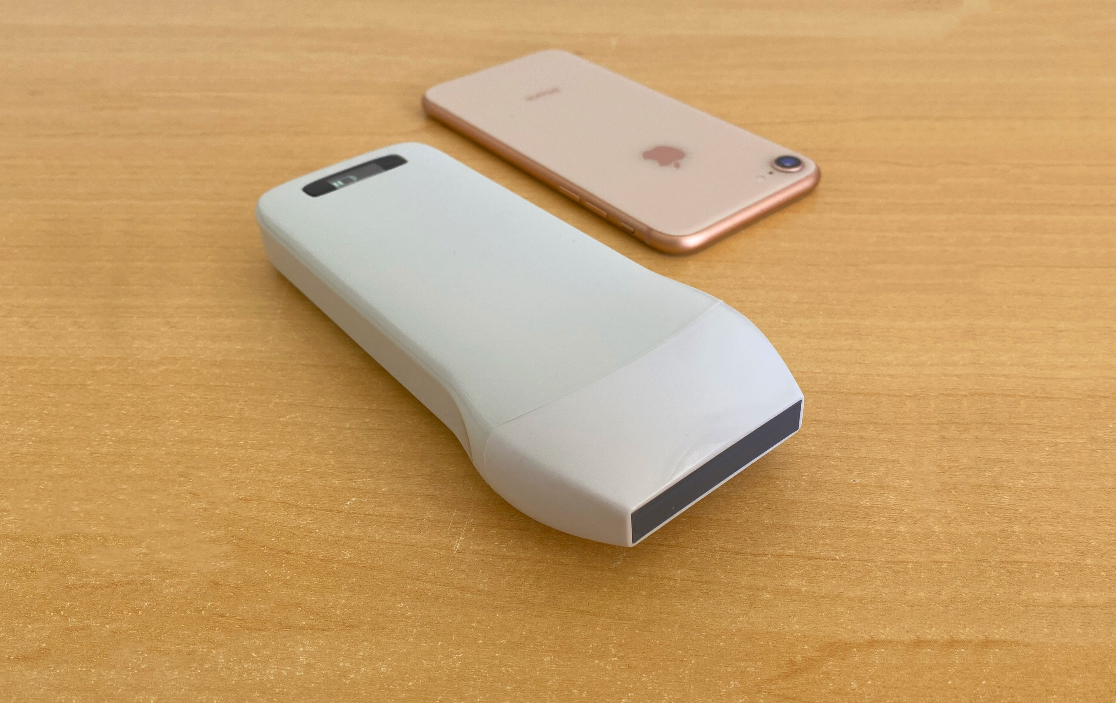
Portable ultrasound devices, such as the C10CX Linear Probe, have revolutionized home-based medical practice. Launched in 2018, this high-definition, wireless device features a scanning frequency of 7.5-10 MHz and 192 elements for rapid, clear imaging. The C10CX is user-friendly and efficient for a range of professionals, including anesthesiologists and physiotherapists. Manufactured by Konted, this portable ultrasound scanner ensures high-quality, real-time diagnostics. Whether for routine monitoring or guided procedures, these devices enable users to deliver high-level care in the comfort of their homes.
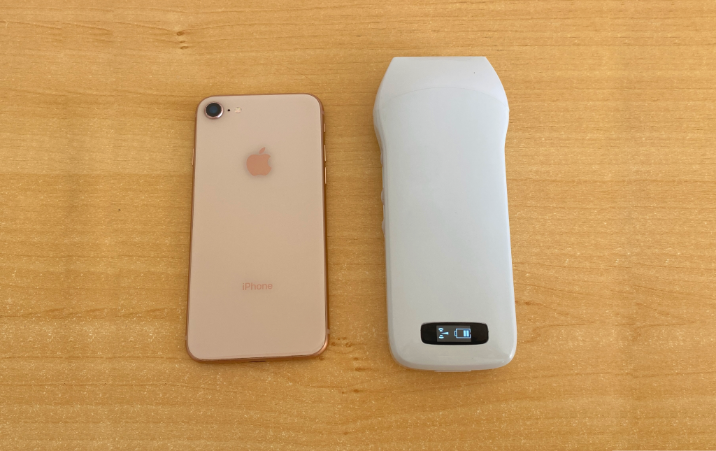
C10CX Color Superficial Ultrasound HD Linear Probe Specifications
| Probe | Wireless linear probe |
| Footprint | L45 |
| Net Weight | 223 g |
| Elements | 192 |
| Frequency | 7.5-10Mhz |
| Depth | 20-80mm |
| Presets | Thyroid, Small parts, Pediatrics, Vascular, Carotid, Breast, MSK, Nerve |
| Host | IOS/Android/Windows, Tablet, Smart phone, PC |
| Connection | WiFi |
| Software Adjustable Parameters | GN(gain), D(Depth), ENH(Enhancement), DR(Dynamic range), F(Frequency), FocusPos, PRF, WF, Mode, 8TGC, Biopsy, Annote |
| Measurement | B: Length, Area, Circumference, Angle, Trace, Distance / B+M: Heart rate, Time, Distance / B+PW: Velocity,Heart rate(2), S/D, Depth |
| Playback Frames | 100, 200, 500, 1000 optional |
| Display Mode | B, B/M, Color, PW, PDI, B+B |
| WiFi Type | Built-in WiFi, 802.11g/20MHz/5G/450Mbps |
| Channel | 64 |
| Frame | 24s/f |
| Gain | 40-110Db |
| Language | Chinese, Ukrainian, German, English, Russian, France, Italian, Spanish, Portuguese (Brazil) |
| Power | Built-in 4200mAh battery |
| Battery Replaceable | Yes |
| Battery Duration | 3 hours (Working time), 12 hours (Stand by time) |
| Charging | Wireless charger, USB cable |
Demonstration Video
Anterior Knee Examination
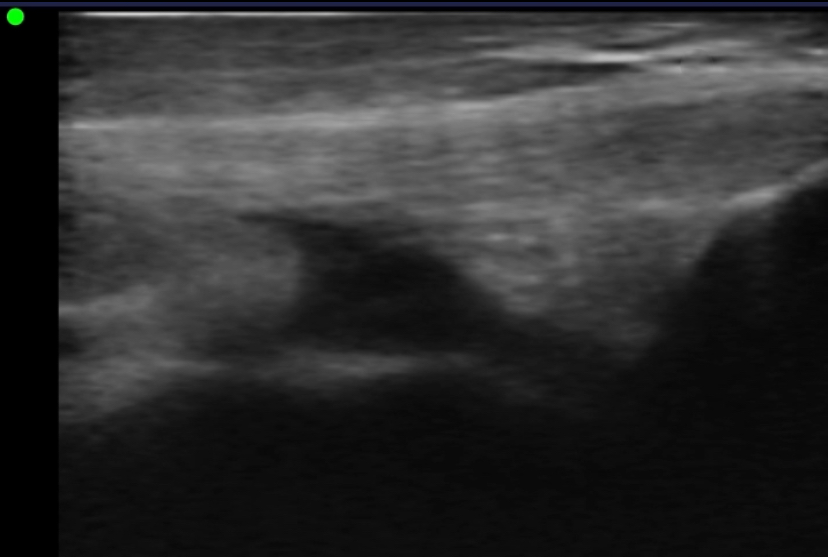
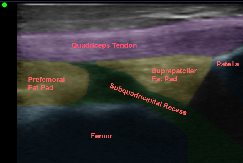
Demonstrated in this video is the use of the C10CX linear ultrasound to locate and identify key anatomical structures in an anterior knee examination of the suprapatellar region. We start with a sagittal examination, highlighting the quadriceps tendon, prefemoral fat pad, suprapatellar fat pad, and suprapatellar bursa. This is followed by a transverse examination to provide a comprehensive view of the suprapatellar bursa and its intra-articular access. This detailed demonstration showcases the capabilities of the C10CX ultrasound in providing clear, high-definition images crucial for accurate diagnosis and treatment planning.
Using Ultrasound for Guided Needle Infiltration
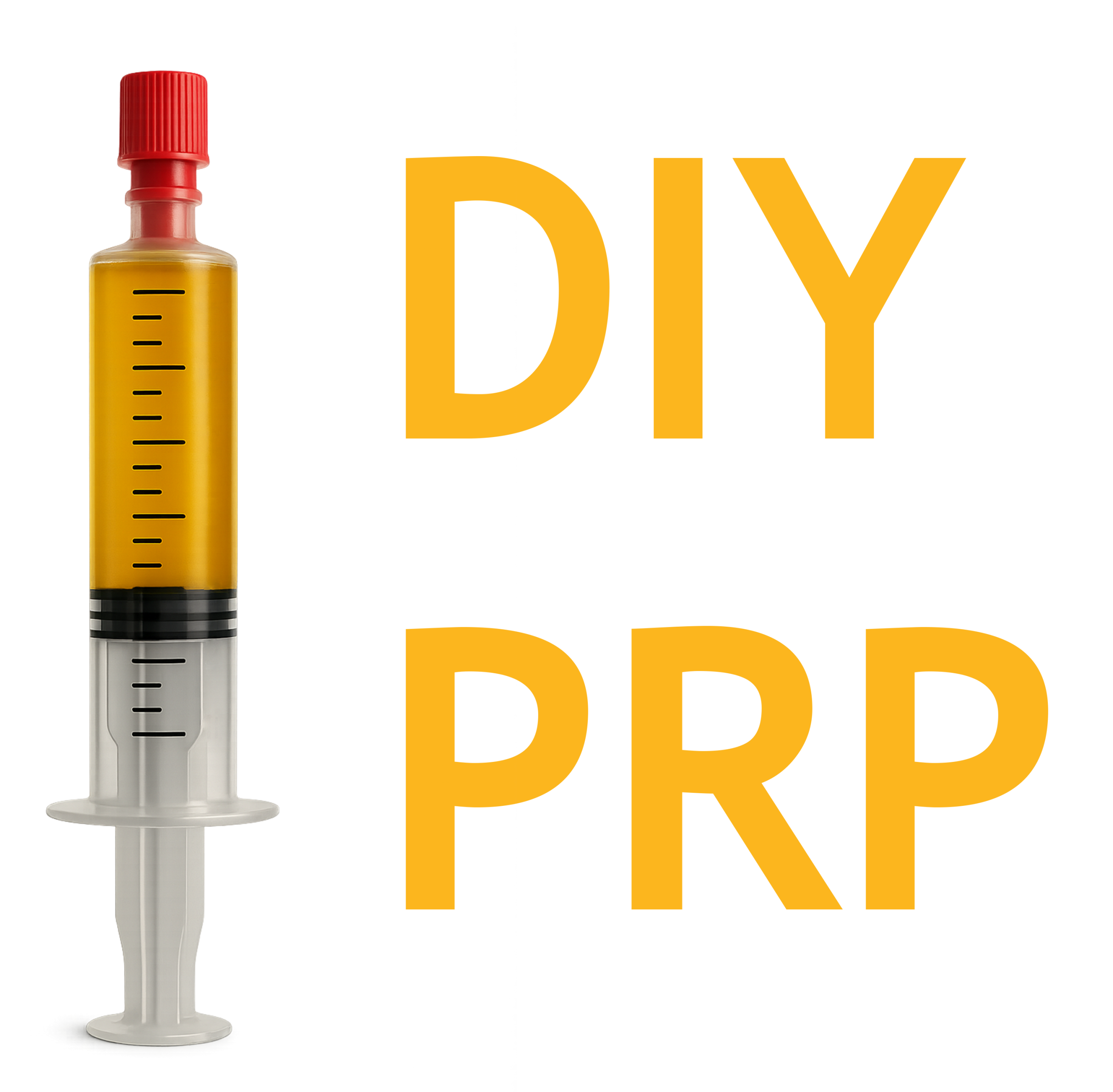
Also demonstrated in the video is needle infiltration. Ultrasound-guided needle infiltration has become a cornerstone in precision medicine, particularly for delivering platelet-rich plasma (PRP) and other therapeutic medications. This technique allows for real-time visualization of the needle path, ensuring accurate placement within the targeted tissue. The enhanced precision reduces the risk of injury to surrounding structures and increases the efficacy of the treatment. Guided injections facilitate better distribution of the therapeutic agents, leading to improved healing outcomes and faster recovery times for patients. The ability to accurately target specific areas within the musculoskeletal system makes ultrasound-guided procedures highly effective for regenerative medicine practices.
Leave a Reply