Preparing Autologous Thrombin Serum from Pre-Isolated PPP for 5 mL PRP Activation
Goal: Produce ~0.5 mL of autologous thrombin serum (ATS) to activate 5.0 mL of PRP.
For instructions on how to formulate a 10% calcium chloride solution (which is required to produce ATS), study our formulation guide beforehand.
Introduction
Autologous Thrombin Serum (ATS) is a powerful, patient-derived reagent created by triggering coagulation in platelet-poor plasma (PPP). The resulting serum is rich in thrombin—a key enzyme in the coagulation cascade—which serves as a potent platelet activator. When mixed with leukocyte-rich PRP (LR-PRP), ATS induces rapid and complete degranulation of platelets, resulting in maximal growth factor release and robust fibrin matrix formation. This makes it particularly ideal for musculoskeletal applications, such as intramuscular or tendon injections, where strong tissue adhesion and rapid regenerative signaling are desired.
While chemical activators like calcium chloride can be used directly to initiate clotting, ATS offers more robust biological activation by mimicking physiological thrombin action. This approach provides enhanced control over clot formation and growth factor kinetics, especially critical in dynamic, high-load environments such as tendons and muscles.
Why and How ATS Is Used in PRP Therapy
ATS is typically used to activate LR-PRP before injection into soft tissues such as muscle or tendon. The thrombin it contains triggers a near-instantaneous platelet activation cascade, ensuring full degranulation and dense fibrin matrix generation at the site of application. This rapid activation profile is especially beneficial when high mechanical forces are present, such as in rotator cuff injuries, hamstring tears, or Achilles tendinopathy.
Compared to direct calcium chloride activation, which relies on slower ionic pathways and fibrin generation, ATS delivers faster, stronger, and more localized clotting, promoting tissue adhesion and a high local concentration of trophic factors. Though more labor-intensive to prepare, its effects are clinically superior for many orthopedic and sports medicine procedures.
Safety Advantages of Autologous Thrombin
The source of thrombin used in regenerative medicine significantly impacts patient safety. Commercially available thrombin products are often derived from bovine plasma, which carries a well-documented risk of immune reactions. These reactions are not solely due to thrombin itself but also to impurities like bovine factor V, which may trigger antibodies that cross-react with human factor V, leading to coagulopathies and potentially life-threatening bleeding events. Additionally, bovine and pooled human thrombin sources raise concerns about transmissible spongiform encephalopathies (TSEs), including variant Creutzfeldt-Jakob disease (vCJD).
For these reasons, autologous thrombin serum, derived from the patient’s own blood, is widely recognized as the safest thrombin source. It eliminates xenogenic and allogenic risks while delivering highly effective platelet activation.
Glass Is Critical
When preparing ATS, the use of glass is not just recommended—it is essential to ensure proper activation of the clotting cascade. The interior surface of glass provides negatively charged silicate groups that facilitate the initiation of the intrinsic pathway of coagulation. This charge is critical for the activation of factor XII (Hageman factor), which sets off a chain of enzymatic reactions ultimately converting prothrombin to thrombin. Plastic, in contrast, is inert and lacks the surface charge required to effectively initiate coagulation in PPP without additional biochemical triggers.
Furthermore, glass enhances thrombin yield by providing a stable surface for fibrin clot formation and does not introduce leachables or hydrophobic interference that might occur with certain plastics. It also improves the visual inspection of clot formation, which is essential in determining the correct resting period before centrifugation. In short, glassware provides both the biochemical surface activity and physical transparency needed for precise and effective ATS generation.
For optimal results, perform all resting and clotting steps in borosilicate or sterilized soda-lime glass containers. Avoid any surface coatings or plastic linings. If transferring to plastic after clot formation, ensure it is a certified medical-grade, low-protein-binding polypropylene or similar material.
Procedure to Create ATS
Materials Required:
- 2.5 mL of pre-isolated platelet-poor plasma (PPP)
- 10% calcium chloride (CaCl₂) solution – sterile, USP grade
- 2 x 4 mL borosilicate tubes (Glass is critical)
- Leur-lock cap
- 2 x 1 mL syringes
- 25G needle
- 22G needle
- Three-way stopcock valve
Instructions:
- Transfer 2.5 mL of pre-isolated platelet-poor plasma (PPP) into a sterile 4 mL glass tube.
- Draw up 0.25 mL of sterile 10% calcium chloride solution from the CaCl₂ vial into a 1 mL syringe using a 25G needle.
- Add the 0.25 mL of sterile 10% calcium chloride solution to the 4 mL borosilicate tube of PPP (maintaining a 1:10 CaCl₂:PPP ratio).
- Gently invert or swirl the container to mix; do not shake.
- Let the mixture within the tube rest on its side at room temperature for 15–30 minutes until a fibrin clot forms.
- While the mixture is resting, make a centrifuge counterweight using another 4 mL borosilicate tube filled with 2.75 mL of water.
- Once the rest time has passed, place both tubes into the centrifuge buckets.
- Centrifuge at 1000g for 5–10 minutes to separate the thrombin-rich serum from the fibrin clot.
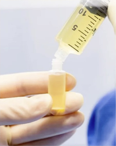
- Using a new 1 mL syringe and 22G needle withdraw approximately 0.5-0.6 mL of the clear supernatant (autologous thrombin serum).
- Remove the needle from the syringe and attach a leur-lock cap.
Note: Use immediately or within 2–4 hours at room temperature under sterile conditions. Do not refrigerate or freeze. The ATS can be transferred to the prepared syringe of PRP using another three-way stopcock valve when ready.
PRP Activation with ATS
- Mix 5.0 mL of PRP with ~0.5 mL of the fresh autologous thrombin serum via a three-way stopcock valve.
- Note: ATS is mixed with PRP in a 1:10 ATS:PRP ratio. Clotting begins in 20 seconds from mixing.
- Using the three-way stopcock valve, manually combine the two mixtures.
- Inject or apply immediately, as clotting begins within seconds.
- Note: The needle should already be positioned in the injection site, or consider using a combination applicator such as a dedicated device or a three-way stopcock valve.
References
- Everts, P. A., et al. (2006). “Platelet-rich plasma and platelet gel: a review.” Journal of Extra-Corporeal Technology, 38(2), 174–187.
– Analyzes the mechanisms of platelet activation, including calcium chloride-induced degranulation and coagulation cascade initiation. - Marx, R. E. (2004). “Platelet-rich plasma: evidence to support its use.” Journal of Oral and Maxillofacial Surgery, 62(4), 489–496.
– Supports the clinical use of calcium chloride in creating autologous thrombin and enhancing tissue adhesion. - Takikawa, M., et al. (2011). “Enhanced effect of platelet-rich plasma containing a new carrier on hair growth.” Dermatologic Surgery, 37(12), 1721–1729.
– Describes calcium chloride-induced thrombin generation in PPP, relevant to enriched serum production like ATS. - Drago, L., et al. (2013). “Platelet-rich plasma for the treatment of orthopedic infections: an in vitro study.” PLoS ONE, 8(9), e73863.
– Demonstrates calcium chloride’s compatibility with sterile and injectable preparations, affirming its utility in PRP workflows. - Mishra, A., et al. (2014). “Platelet-rich plasma in orthopedic applications: evidence-based recommendations for treatment.” Journal of the American Academy of Orthopaedic Surgeons, 22(18), 469–470.
– Outlines PRP applications in tendon and muscle injuries and the importance of controlled activation. - Burnouf, T., Strunk, D., Koh, M. B. C., & Schallmoser, K. (2016). “Human platelet lysate: Replacing fetal bovine serum as a gold standard for human cell propagation?” Biologicals, 44(5), 387–400. https://doi.org/10.1016/j.biologicals.2016.01.010
– Explains the immunologic risks of bovine thrombin and rationale for autologous sources. - Kumar, V., & Chapman, J. R. (2008). “Autologous Thrombin: Intraoperative Production From Whole Blood.” Techniques in Orthopaedics, 23(2), 116–119. PMCID: PMC4680638
– Reviews clinical technique and rationale for autologous thrombin production.
Leave a Reply